Medical Imaging Analysis
How computer vision and deep learning help detect diseases from X‑rays, CT scans, and MRIs.
What is Medical Imaging Analysis?
Medical imaging analysis uses machine learning algorithms to automatically interpret images such as X‑rays, CT scans, MRIs, and ultrasounds. In a triage setting, these systems can flag critical findings (e.g., a collapsed lung, brain bleed, or bone fracture) within seconds, helping clinicians prioritise the most urgent cases.
1. Key Computer Vision Tasks
Modern medical image analysis typically uses convolutional neural networks (CNNs) – deep learning architectures specially designed for grid‑like data (images). Common tasks include:
- Classification: Does this image contain a disease? (e.g., “pneumonia present” / “normal”).
- Segmentation: Outline the exact region of disease (e.g., highlight the tumour boundary).
- Detection: Locate and label multiple abnormalities in one image (e.g., all rib fractures).
- Quantification: Measure disease burden (e.g., percentage of lung affected by pneumonia).
These outputs become part of the structured data that feeds into the triage engine – a positive finding can increase the probability of a critical condition.
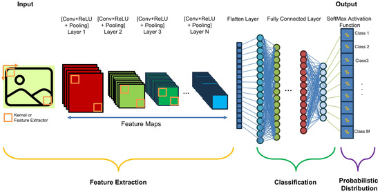 Figure: A convolutional neural network (CNN) processes the image layer by layer to make a prediction.
2. Integration into the Triage Workflow
A typical real‑time medical imaging triage pipeline consists of these steps:
- Image acquisition: DICOM (Digital Imaging and Communications in Medicine) images are sent from the scanner to a PACS (picture archiving and communication system).
- Machine learning inference: A trained computer vision model (e.g., deep learning network) automatically analyses the images – detecting abnormalities, segmenting lesions, or classifying findings. The exact architecture may vary (CNNs, Vision Transformers, or ensemble methods) depending on the imaging modality and clinical task.
- Structured output generation: The model's results are converted into a structured vector of findings (e.g., `pneumothorax: true`, `location: right lung`, `confidence: 0.94`). This vector becomes one input to the broader triage engine (alongside symptoms, lab results, etc.).
- Triage integration and alerting: The triage engine combines imaging findings with other clinical data. If the combined assessment crosses a critical threshold, the dashboard shows a priority alert – guiding the clinician to review the most urgent cases first.
 Figure: The AI pipeline integrates seamlessly into existing radiology workflows – alerting clinicians when urgent findings are detected.
3. Challenges in Veterinary Imaging AI
Data annotation: Labelling medical images requires expert veterinary radiologists – expensive and time‑consuming, especially across multiple species.
Generalisation: A model trained on images from one clinic's scanners may fail on images from a different manufacturer or animal population (e.g., different breeds, sizes).
Explainability: “Black box” models are hard to trust in high‑stakes settings. Techniques like saliency maps or attention heatmaps help clinicians understand the model's focus.
Regulatory approval: Clinical deployment may require veterinary‑specific certifications (e.g., CE‑mark for veterinary devices, or local board approvals) – a lengthy process.
Integration: Seamless interaction with practice management software, PACS, and electronic medical records (EMR) is non‑trivial.
Bottom line: Veterinary imaging AI requires careful validation across species, regulatory attention, and workflow integration – but the potential to improve triage speed and accuracy is substantial.